
Compare the conditions before comparing the contours. Fat-freezing before-and-after photographs are most useful when the camera, lighting, pose, clothing and view match, and the dates and treatment details are clear. Even then, they are one part of an assessment: a photograph records an appearance, not the amount of fat removed or the reason a shape changed.
For your own record, aim for a small, repeatable set of images that you can discuss at review. There is no need to photograph yourself every day or recreate someone else’s advertised result.

Decide what you are trying to assess
Cryolipolysis, or fat freezing, targets suitable local fat beneath the skin. It is not a weight-loss treatment. That distinction matters when choosing evidence: bathroom scales measure total body weight, not the contour of the treated area. The FDA’s body-contouring guidance explicitly separates these procedures from weight loss and its health benefits.
An unchanged weight therefore does not settle whether there has been a local result. Conversely, if your weight falls between photographs, the pictures cannot separate the contribution of that change from the procedure. Keep weight information as context if available, rather than setting a kilogram target for treatment success.
Before treatment, describe the particular change you hope to see: perhaps the outline of one flank, rather than a generally smaller waist. Ask which views would show that area and what would count as a meaningful, realistic improvement. Keep the same question for the follow-up discussion.
Look for comparable photographs
Published medical-photography guidance explains that changes in camera distance, angle, illumination and posture can alter an apparent result. Even breathing can change the appearance of the abdomen. These are photography principles, not evidence that a particular treatment has worked. See the proposal for photographic documentation standards.
Cryolipolysis researchers have taken similar precautions. In a primary flank study, photographs used fixed foot and arm positions, consistent equipment and lighting, and independent reviewers who were not told which image came first. This illustrates how much more controlled a research comparison can be than two casual selfies; it does not make that study’s method a compulsory protocol for every clinic. Read the study’s photographic evaluation method.
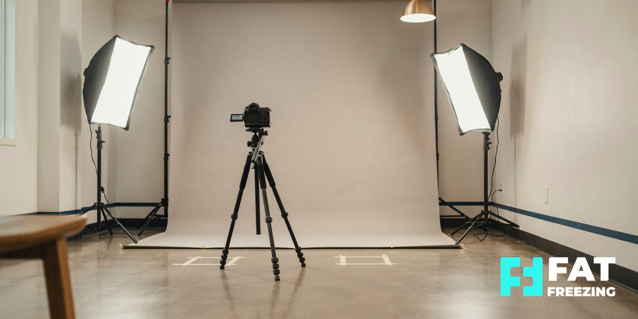
Use this practical checklist when inspecting a gallery or planning your own pictures. It translates those principles into questions; it is not a validated scoring system.
| Check | What makes comparison more useful? |
|---|---|
| Light and background | The same background and light direction, without new shadows emphasising the outline. |
| Camera and distance | The same camera or phone lens, height, distance and framing; no switch to a close wide-angle selfie. |
| Pose and breathing | Matching feet, arms and body angle, with a relaxed posture; no deliberate sucking in or twisting. |
| Clothing | The same garments in the same position, without different waistbands compressing or hiding the area. |
| Views and editing | Matching front, side or angled views as appropriate; original images without body reshaping or beauty filters. |
| Dates and context | Dates, treated areas, number of sessions, other procedures and relevant weight changes identified. |
If a detail is missing, ask for it. A dramatic difference under unmatched conditions is a reason to reserve judgement, not proof that an image is dishonest. Equally, a polished gallery may show selected examples; it cannot tell you your own likelihood of achieving the same result.
Keep a manageable record over time
The following is an organisational suggestion, rather than a published clinical measurement schedule: keep a dated baseline set, a short note of the setup and a record at the review interval agreed with your practitioner. A floor marker for the camera and another for your feet may help you repeat the arrangement. Store originals together and label the body view clearly.
Note circumstances that made a session unusual, such as noticeable bloating, different clothing or a changed camera. Keeping broadly similar everyday conditions is useful; do not change eating or drinking simply to produce a more flattering photograph. If the setup cannot be matched, record that limitation instead of editing the image to compensate.
Timing needs context too. The American Academy of Dermatology describes gradual results from non-invasive fat removal and temporary swelling, bruising or discomfort. An early image may therefore document recovery rather than the eventual contour. Agree appropriate review timing for your device and treatment plan; our results timeline guide explores this separately.
Keep personal images private and ask how any clinic photographs will be stored and used. Discuss permission for clinical records separately from permission to publish pictures in marketing.
What can a tape measure add?
Circumference means the distance around a body area. It can supplement photographs, but it does not isolate the treated fat layer. A slightly different tape level, angle or tightness makes the comparison less dependable.
A primary inner-thigh cryolipolysis study illustrates the care involved: investigators recorded the measurement height, took each circumference three times and averaged the readings. They also used photographs and ultrasound. That was a method for the particular study site and device, not a universal waist-measuring rule or a home test of effectiveness. Read the inner-thigh study.
If you want to keep measurements, ask the practitioner to demonstrate a repeatable method appropriate to your area. Record the method as well as the number. Treat small differences cautiously, especially if you cannot reproduce the reading comfortably. Do not translate centimetres around the waist into a percentage of fat removed.
Bring the record to a clinical review
Use the images to explain what you notice and ask whether examination supports that impression. If there is little change, discuss the limits of the comparison and the treatment response before assuming another session is the answer. A later change raises a different question; see how long fat-freezing results last.
A record must not delay care. Seek assessment for severe or worsening pain, blistering, marked colour change or a growing firm bulge. Paradoxical adipose hyperplasia is uncommon enlargement and hardening of treated fat that can appear months later, does not normally resolve by itself and may require surgery. Photographs cannot diagnose it. FDA information on cryolipolysis complications.
For a Nottingham consultation, bring your questions about documentation and review to the clinic at Atomic House, 50 High Pavement, or contact the team before attending.
Sources and further reading
- FDA: Non-Invasive Body Contouring Technologies.
- American Academy of Dermatology: Non-invasive fat removal — what can you expect?.
- Prantl and colleagues, 2017: A Proposal for Updated Standards of Photographic Documentation in Aesthetic Medicine.
- Non-invasive fat reduction of the flanks using a new cryolipolysis applicator and overlapping, two-cycle treatments.
- Cryolipolysis for safe and effective inner thigh fat reduction.
Frequently Asked Questions
Can a before-and-after photo prove that fat freezing worked?
A well-matched pair can document a visible change, but cannot by itself establish its cause. Lighting, posture, clothing, weight changes and other treatments need to be considered alongside timing and clinical assessment.
Should I use my weight to judge a local result?
Weight provides context, but fat freezing is intended for local contouring rather than weight loss. An unchanged scale reading does not rule out a local change, and a lower reading does not establish that cooling caused it.
Are home circumference measurements worth keeping?
They may provide supporting information if you can repeat the same method comfortably. Tape position and tension affect the reading, and circumference is not a direct measurement of the treated fat layer. Ask the practitioner how they assess the particular area.
What if the treated area looks larger in later photographs?
Arrange an assessment rather than diagnosing the change from photographs. A growing firm bulge months after treatment can be a sign of paradoxical adipose hyperplasia, an uncommon complication that does not normally resolve by itself and may require surgery.

